USA & Canada
USA & Canada
Trainee surgeon using a 3D printed model of a patient’s kidney in the operating theatre during a surgery.
Case Study

Bordeaux University Hospital in France enhances kidney operations with full-color, multi-material 3D printing.

October 01, 2019

Taking a new approach with 3D printing technology.

The powerful impact of 3D printing on health care is evident across leading hospitals throughout the world. Bordeaux University Hospital (CHU), France, is one notable example where this is apparent. As one of the first hospitals worldwide to adopt full-color, multimaterial 3D printing using the Stratasys J750 3D™ printer, its surgical teams are able to benefit from its broad capabilities. These range from advancing surgical processes using ultra-realistic pre-surgery planning models to improving student training and enhancing patient education.

Spearheading the use of 3D printing is the Department of Implantology, Urology and Kidney Transplantation, which pioneered its deployment to aid with complicated kidney tumor removal. During that time, the team of physicians, led by CHU surgeon Professor Jean-Christophe Bernhard, has performed a number of operations involving the use of transparent and color 3D printed models. The CHU is producing complex patient-specific models that mix transparency with multiple colors to visualize different structures within the anatomy. The transparent model shows the volume mass of the kidney itself with red elements for the arteries, blue for the veins, yellow for the excretory tract and pink for the tumor. The entire model is also produced in a single print operation..

Having a 3D printed kidney model in your hands that corresponds specifically to that of the patient you’re going to operate on offers us a view from a new perspective.

itw01263203still026 high res

More precise and successful kidney surgery.

According to Prof. Bernhard, the clearer view offered by the 3D printed model increases the ability to perform precise and successful kidney-sparing surgery under super selective clamping. The model aids with pre-surgery planning, identifying the delicate nearby arteries and vessels that can result in complete kidney removal if damaged.

“A scan gives us good information, but it’s in 2D,” said Prof. Bernhard. “This relies on the surgeon to mentally reconstruct the tumor volume in 3D and estimate its location inside of the total volume of the kidney. The same process has to be done to clearly understand the relations between the tumor, the vessels and the collecting system. As you can imagine, this is difficult and time-consuming for the surgeon.

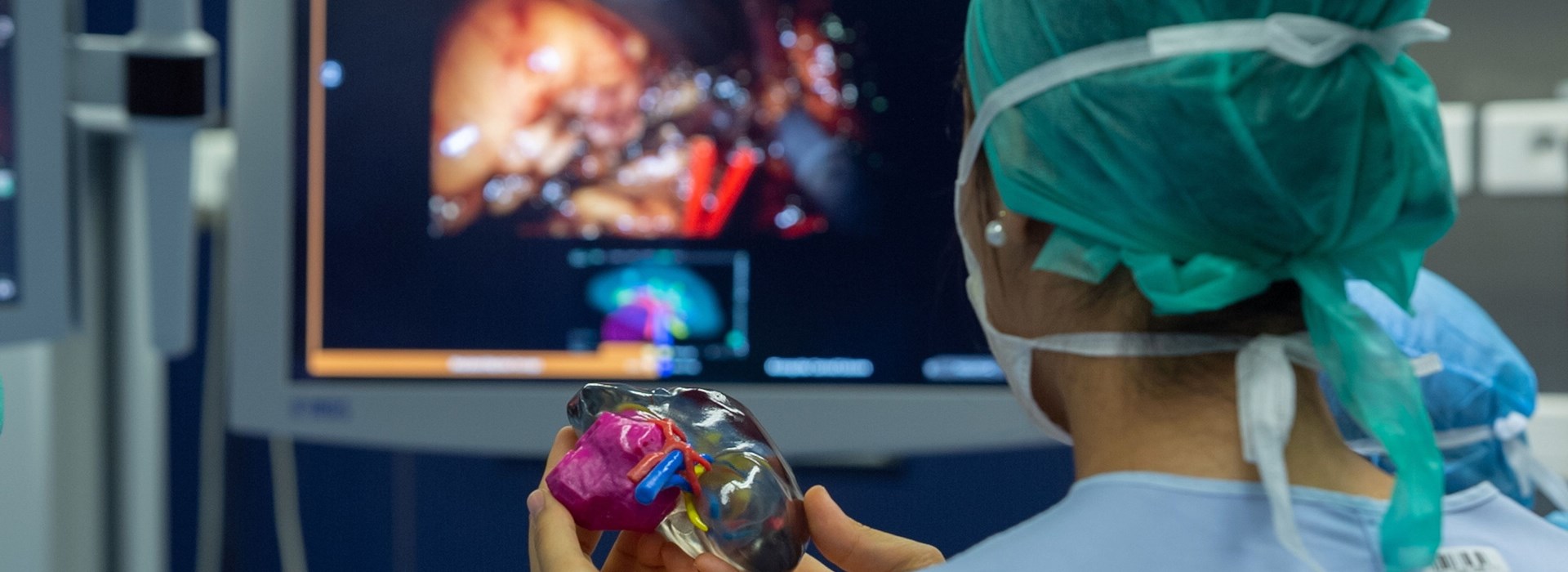
“Conversely, having a 3D printed kidney model in your hands that corresponds specifically to that of the patient you’re going to operate on, quite literally, offers us a view from a new perspective. The only thing more accurate than that is the patient himself,” he added.

Prof. Bernhard initiated a research project entitled “Rein 3D Print.” The study aims to improve the communication process with patients and assess their satisfaction with 3D printed models for pre-therapeutic education. Moreover, boosting patient understanding of their surgical procedure can potentially increase the possibilities of ambulatory care (same-day surgery that doesn’t necessitate an overnight stay). Crucial to the project is the J750, which was acquired from Stratasys reseller and funded by the European Union and the Regional Council of France’s Nouvelle Aquitaine region.

Enhancing patient understanding and care.

“Describing kidney tumor removal with a 2D scan or drawing will invariably leave most patients somewhat bewildered,” explained Prof. Bernhard. “Presenting them with a 3D printed model that clearly shows the tumor puts them at ease and enables the patient to grasp exactly what we’re going to do. Indeed, initial research from patient questionnaires shows that having 3D printed models increases their understanding of the surgery by up to 50%, so it’s a considerable benefit in terms of overall patient care.”
Unique color, multi-material 3D printing enables CHU to 3D
print ultra-realistic models of patients’ kidneys, providing clear
visualization of tumors (pink area) pre-surgery.
Trainee surgeon using a 3D printed model of a patient’s kidney in the operating theatre during a surgery.

Improving instructional techniques for tomorrow’s surgeons.

As Prof. Bernhard explained, another key objective of the “Rein 3D Print” project is to constantly improve instructional techniques for trainee surgeons. “Having access to a 3D printed model that is completely accurate to the one that you’re going to operate on enables you to train yourself on the operation. Additionally, it also greatly improves our ability to more accurately convey surgical procedures to students,” he said.

The J750 is operated by a team at the Bordeaux IUT fablab unit called Coh@bit, which finalizes virtual models of patients’ kidneys and develops STL files from these images. The models are then 3D printed and delivered to the CHU. In some instances, the whole process is completed within a day. Having previously outsourced its 3D printed models, the CHU has shortened lead times and eradicated freight and other external costs since installing the J750, allowing the medical team to be far more reactive. This means surgeons can plan operations and treat patients within a much shorter timeframe. Communication between the CHU medical teams and the IUT technical teams has also been strengthened. According to Prof. Bernhard, the J750 3D printer has facilitated interactions, creating and encouraging knowledge sharing and ideas.

Download the case study

Download