Children’s Hospital Colorado, a leader in pediatric heart care, is improving the lives of children with complex congenital heart disease with the help of patient-specific 3D models. As a referral center, it sees some of the most complicated congenital heart defects. One such case was a 4-year-old child born with tricuspid atresia, acongenital heart defect that affects approximately 1 in 10,000 births in the United States. Children with this condition are born with low oxygen concentrations in the arterial blood and are referred to as cyanotic. A series of palliative surgeries are typically undertaken to restore a more normal oxygen saturation level and improve life expectancy and quality of life. For Children’s Hospital Colorado, this defect is not that rare. It successfully treats many children with tricuspid atresia each year. However, this particular case presented a unique challenge because it involved an extremely rare variant in which the ventricular cardiac chambers were arranged in a superior-inferior position compared to the typical side-by-side arrangement.The mass or apex of the heart was pointed to the right instead of its normal left position.
The great arteries were oriented opposite their usual relationship to each other and both originated from the upper small right ventricle. The right ventricle wassupplied from the left ventricle through a large hole called a ventricular septal defect. The smaller atrial chambers that collect blood coming back to the heart were rotated posteriorly and to the left, resulting in a plane of division that was much more transverse than usual. This highly unusual anatomy was also complicated by Wolff-Parkinson-White (WPW) syndrome. This condition involves an accessory electrical pathway between the ventricular and atrial chambers that leads to dangerously fast heart rates. In a patient with only one functional ventricle, WPW syndrome is extremely perilous and if unaddressed, increases the risk of death following cardiac surgery. The patient had undergone two previous palliative surgeries but because of the discovery of WPW syndrome, she was referred to the Heart Institute at Children's Hospital Colorado.
Max Mitchell, M.D., a congenital cardiac surgeon,and Kathryn Collins, M.D., a pediatric cardiologist specializing in electrophysiology, reviewed the child’s history to determine her next treatment steps. Atbirth, her tricuspid valve that normally controls blood flow from the right atrium to the right ventricle was not formed, preventing blue blood from getting to her right ventricle and out to her lungs. The main pulmonary artery was also too small to provide adequate blood supply to her lungs. A hole between the right and left atria, known as an atrial septaldefect, allowed a mix of oxygen-rich and oxygen-poor blood to be pumped through her body. To address these issues and enable the child to grow, a shunt had been placed in her as a neonate between the pulmonary artery and the aorta allowing blood to get to the lungs. And at 12 months of age, she underwent a bi-directional Glenn procedure, the first in a two-stage surgical strategy that by passes the heart entirely and pumps blood returning from the tissues directly to the lungs. After the Glenn procedure, Dr. Mitchell and his colleagues determined that the child was a candidate for the next stage of surgery known as the Fontan procedure, the second in the two-stage surgery.
However, a successful Fontan procedure is highly dependent on a normal heart rhythm and heart rate.The arrhythmia associated with WPW syndromehad to be addressed either prior to surgery in the catheterization lab or at the time of surgery. In addition, it was imperative that her accessory electrical pathway be ablated prior to completionof the surgery because it would be impossible to address in a catheterization laboratory after the Fontan procedure, and because all the venous connections from her body to her heart would no longer exist. This would leave no route to get a catheter to the accessory pathway. Dr. Collins attempted to ablate the accessory pathway in the catheterization laboratory but was unable to contact the accessory pathway due to the markedly abnormal anatomy. As a result, she approached Dr.Mitchell to gauge the feasibility of a surgical ablation at the time of the planned Fontan operation.
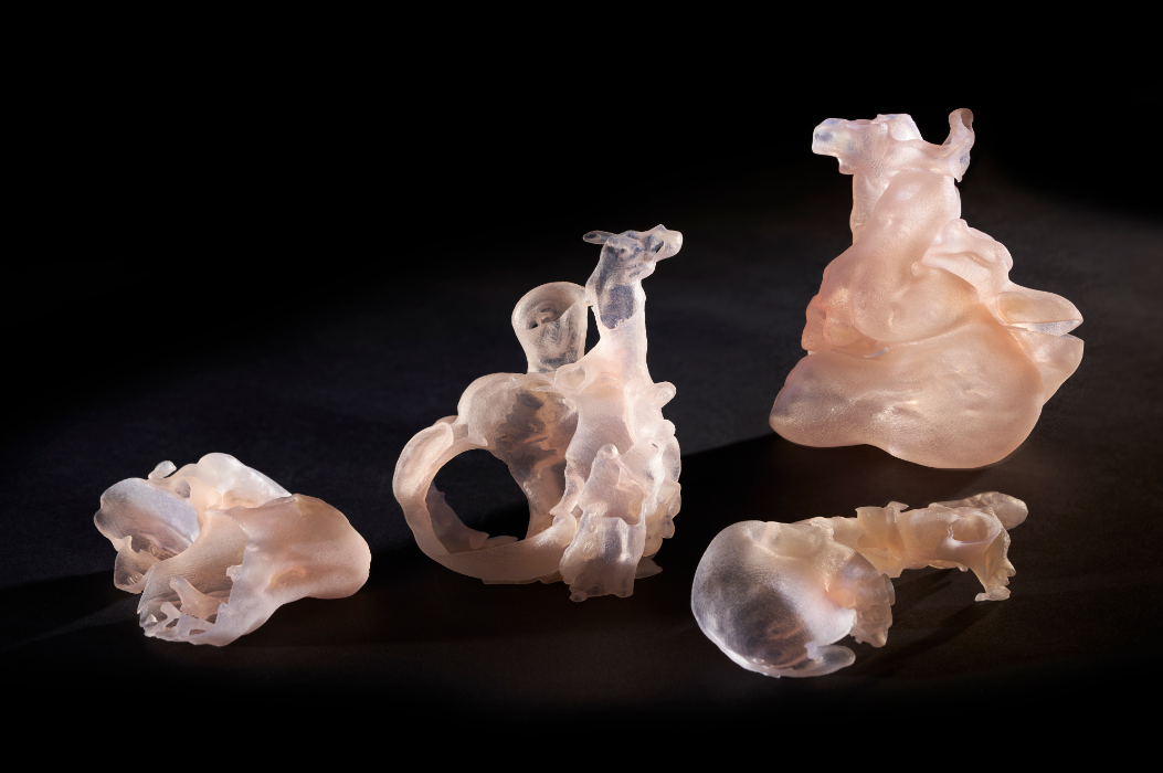
Even for experienced congenital heart surgeons, echocardiograms are not easily related to 3D anatomy in complex, rare cardiac anomalies.After reviewing an MRI scan of the patient’s heart with 3D reconstructions, Dr. Mitchell was concerned that a surgical ablation carried a high risk of failure. He approached colleagues at the University of Colorado Anschutz Medical Campus to have a 3D patient-specific model printed and cut into sections for a better understanding of her cardiac anatomy.The model needed to be printed with materials that simulated the look and feel of the patient’s heart. The hope was that this would guide a successful second attempt at ablation in the catheterization lab.Lorna Browne, M.D., the lead radiologist on the case,prepared the scans and teamed up with designers atIn works, a design studio at the University of Colorado Anschutz, to create the 3D model.
Nicholas Jacobson, the design lead on the case, and Hayden McClain, student assistant, helped design the 3D printing process for pre surgical planning with the team — orchestrating early brain storms to determine the design of the model that would be used. Dr. Michael DiMaria, lead cardiologist on the case, was instrumental in guiding Jacobson in determining which anatomy was crucial to capture in the model due to his intimate knowledge of the patient’s diagnosis. Designed to fit the team’ssurgical specifications, the model was ultimately created using a custom Agilus30™ and Vero™ White multi material blend.In preparing for the electrophysiology (EP) labprocedure, Dr. Mitchell and the cardiology team used the model to plan the approach. The flexible and functional nature of the model allowed them to stretch and pull certain areas to understand the interior topology and flow paths. The EP team used the MRI overlaid on the lab’s intra cardiac navigation system with concurrent real time transesophageal echocardiography.
“What I learned from the model had a big impact not only on our ability to successfully complete the ablation but also on my surgical approach. The way things laid out in surgery was exactly what I expected based on the model. It gave me an accurate understanding of all the anatomical relationships,including the mal position of the great arteries and the enlarged aorta that arose from the unusual position of the small right ventricle,” Dr. Mitchell added.
“I did the operation differently from my usual approach based on the information the model provided. Holding the model in my hand allowed me to visualize what I would see in surgery and from that I was able to plan a more informed approach,which allowed me to go into the surgery with more confidence. The model also allowed me to design a more efficient Fontan for her and do it safely through all of the scar tissue that was there from previous surgeries.”
Dr. Mitchell said, “With that model and having it cut into the appropriate planes, we were able to go into the EP lab with our electrophysiology and echocardiography teams and pinpoint the catheter location inside the child’s heart within millimeters.Using their navigation system, the echocardiogramand the models, all simultaneously, we were all able to stay oriented during the ablation and were visually able to anticipate the pathway to the suspected location of the accessory pathway. This facilitated Dr.Collins’ ability to guide the catheter to the accessory pathway and achieve a successful ablation.”
To further assess the movement of blood through her heart, Dr. Mitchell’s research team performed a 4D-MRI flow analysis.
This allows the investigator to picture the blood flow through the heart and into the blood vessels. Due to the very unusual orientation of the takeoff of her aorta, Dr. Mitchell suspected that blood flow through the ventricles and into the aorta would be highly abnormal and likely inefficient. This proved correct, and he requested a multimaterial 3D print of the heart demonstrating the very unusual blood flow pathways prior to surgery.
Dr. Alex Barker, the Director of the Advanced Imaging Lab and early pioneer of the 4D Flow MRI imaging process, worked with Dr. Mitchell and Jacobson to generate and extract the 4D flow data into a format that could be used to 3D print the new heart model. Each color used in the model was coded to a velocity.
“The model provided comprehensive flow hemo dynamic information ateach anatomic region throughout the cardiac cycle.It demonstrated that everything about the flow in her heart was highly abnormal and confirmed thather pumping efficiency would likely remain more inefficient after the Fontan procedure comparedto patients with more typical tricuspid atresia. This advanced 4D-Flow MRI analysis model provided valuable information to direct future potential management changes,” stated Dr. Mitchell.
Ultimately, both the surgeon and the patient benefited from this personalized approach to medical treatment.
Dr. Mitchell summed it up, “I gave her the best-informed surgical treatment possiblethanks to the model and she continues to do well post-operatively.”


