One of the key elements to educating and training healthcare practitioners is understanding both normal and pathologic anatomy. In the real world, every patient’s anatomy is different, so a surgeon’s practice on human cadavers, animal models and generic mannequins often has little relevance to the actual patient on the table. Existing training models have significant limitations. Human cadavers are in short supply, provide a limited pathology range and are rarely matched to the target pathology in training. Plus, they do not retain the responsiveness of living tissue. Animal models are instructive on the principles of surgery (e.g., cutting, suturing, deploying or attaching devices), but do not replicate human anatomy.
Both animal and cadaver training models require expensive training and controlled environments. Mannequins are limited to a recreating normal or generic anatomy due to the high cost of producing variations and the limitations of tooling used to mass-produce these mannequins. Of course, a surgeon can study the 3D shape of the patient’s anatomy on a computer screen using images acquired from magnetic resonance imaging (MRI) and computed tomography (CT). But developing surgery skills requires physically interacting with a full range of complexity in human pathology.
With advancing 3D printing technology, creating realistic examples of human anatomy is not as time consuming or costly as traditional methods of medical modeling. So it is not surprising 3D printed medical models are increasingly used by surgeons and more often the focus of studies published in medical literature. Historically, 3D printing required trade-offs because of the limited physical properties and color capabilities of systems developed for rapid prototyping. But with more than 360,000 colors and textures as soft as tissue and as hard as bone, the Stratasys J750 3D printer enables the unparalleled ability to recreate human anatomy in a clinically relevant training simulator.
“The Stratasys J750 can create models of individual patients’ anatomy so surgeons can plan out the best way to perform the surgery, and practice the procedure over and over prior to entering the operating room,” said Crispin Weinberg, president of Biomedical Modeling Inc. (BMI), a leading producer of medical models. BMI engineers used medical processing and CAD software to separate (commonly referred to as segmentation) MRI and CT DICOM data of actual patients into different types of tissue. Post-processing software reverse engineered, refined and converted the geometry into a surface model. The BMI team used graphical software to add color, transparency and flexibility information to the model and generated a VRML file to print.
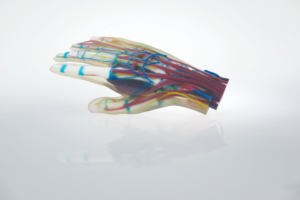
BMI created an unprecedented model of a human heart with the Stratasys J750 using photographs of operations and cadavers to guide the segmentation of the model and application of realistic colors. The original design was a single solid heart, but a quick modification allowed it to be printed in two parts, allowing trainees to remove the front and view internal chambers, valves, papillary muscles and other structures. BMI also used the Stratasys J750 to create a cross-section of a head and brain for anatomical education and technical demonstrations. It uses the same materials as the heart but instead of color realism, vibrant and differentiated colors highlight different biological tissues while transparent material reveals internal structures and tissues. BMI created a hand model that also provides realistic colors and uses a transparent and flexible material on the model’s skin layer to provide a realistic texture and transparency to see the critical structures underneath.
“The Stratasys J750 can economically produce training mannequins in a wide range of normal and abnormal anatomies, replicating realistic colors and tissue textures,” Weinberg said. “The 3D printer makes it possible for surgeons to effectively train on abnormal and patient-specific anatomies.”
3D printing is ushering in a new age in medical anatomical models with a degree of complexity, realism and sophistication not previously possible. Designers can economically recreate any anatomy with texture and color realism. For the first time, it is practical for surgeons to practice on a wide range of abnormal or patient-specific anatomy.

