Brain lesions and tumors are generally identified through magnetic resonance imaging (MRI) or computed tomography (CT) scans, but these modalities may be limiting in providing eloquent details related to these conditions. That’s why Dr. Darin Okuda, Director of Neuroinnovation at UT Southwestern, is utilizing 3D printing to reproduce brain lesions resulting from multiple sclerosis (MS), an autoimmune disease that causes neurodegeneration of the central nervous system and acute inflammatory attacks, and tumors caused by glioblastoma multiforme (GBM), an aggressive cancer that occurs in the spinal cord or brain.
The study of the physical 3D models has allowed Okuda and his team to appreciate the qualitative characteristics of disease that are not apparent in orthographic view.1 “This may be the result of the impact of light coming into the model at different angles allowing for a deeper appreciation of the characteristics present. Our visual inspection of the physical models has also allowed us to develop new hypotheses that we have incorporated into our machine and deep learning systems,” says Okuda.
With the use of 3D printing, Okuda and his team have been able to better understand brain lesions related to MS and tumor growth/regression which has led to greater insights into disease evolution as well as improving treatment and patient compliance and care.
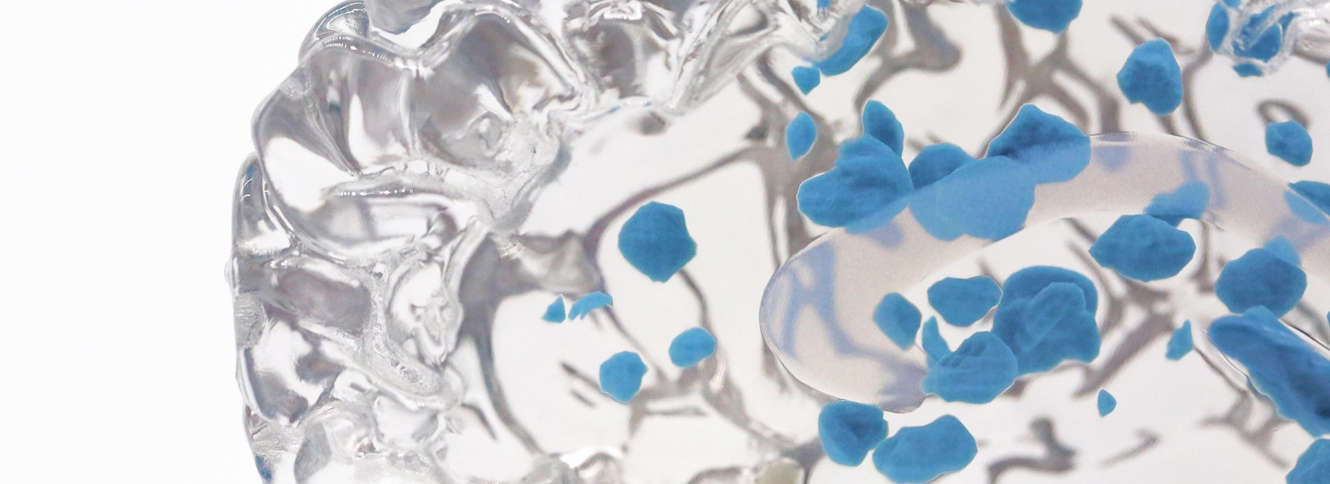
A 3D printed model is more effective than a twodimensional image that is limited in depicting the intricacies of a lesion from MS within the brain.
“The true shape of a lesion within the brain is nearly impossible to characterize when viewing images in a forced perspective, two-dimensional plane,” Okuda explains. “By studying lesions in 3D, we are looking at these findings in an entirely different way — assessing their shape, structure and surface characteristics.”
3D printing allows for the appreciation of differences in physical properties between lesions. Using this medium, the unique characteristics of each lesion can be appreciated, highlighting differences in lesion age, degree of injury, as well as potential for recovery and repair.
In addition, 3D printing allows for new scientific hypotheses for future research. For example, the 3D models allowed Okuda and his team to hypothesize that surface textures may be different between race and ethnicity in MS. And indeed, when they compared the “dorsal sections of the medulla-upper cervical spinal cord” of African American (AA) and white patients, they “identified greater local surface texture changes involving the anatomical regions containing the cuneate and gracile nuclei, nucleus of the solitary tract, medial vestibular nucleus, and hypoglossal nucleus when AA and white MS patients, without any signs of neurological impairment, were compared”.2
Leveraging 3D printing has led to a different perspective in appreciating the relationship between the underlying biology of disease and physical structure.2, 3, 4, 5
To treat GBM successfully, it’s crucial to view the tumor growth accurately.6 However, 2D imaging limits the understanding of tumor growth and doesn’t reveal the shape and texture. And, as a patient, information from CT scans and MRI studies can be confusing.
With the use of 3D models, doctors are not only better able to understand the structural differences that inform disease advancement or regression, but it allows them to educate patients more intuitively by providing tangible data.
“Seeing medical conditions printed in true physical form improves how we educate patients and provides an immediately intuitive platform for the understanding of disease,” says Okuda. “The ability to understand what’s happening inside of you, to actually hold it in your hands and see the effects of treatment, is incredibly empowering.”
When doctors utilize 3D printing models, it can really make an impact because a patient can hold their own tumor in their hand. Not only does a model provide more clarity, but it also helps patients to understand the rationale for the provided medical recommendations, as it offers a physical connection with what is happening in the body, which often causes a patient to follow their treatment plan more carefully.
Okuda used PolyJet™ Digital Materials from Stratasys to print his models because with multi-color and multi-material capabilities with microscopic layer resolution and accuracy down to 0.014 mm, PolyJet prints the most patient-specific models in intricate detail with unlimited possibilities to choose colors, define transparencies and determine textures and finishes.
“In working with Stratasys’ VeroUltra™Clear Transparent PolyJet Digital Material, we now have the flexibility of printing professional grade brain and spinal models to physically demonstrate transitions in disease states, a facet never before available within the 3D printing space,” says Okuda.
Okuda concludes that utilizing 3D printing “provides an innovative solution to not only better understand the nature of problems in the neurosciences but transforms the delivery of education to healthcare providers and the patients we serve.”
1 Newton BD, Wright K, Winkler MD, et al. Three-Dimensional Shape and Surface Features Distinguish Multiple Sclerosis Lesions from Nonspecific White Matter Disease. J Neuroimaging 2017;27:613-619.
2 Moog TM, McCreary M, Stanley T, et al. African Americans experience disproportionate neurodegenerative changes in the medulla and upper cervical spinal cord in early multiple sclerosis. Mult Scler Relat Disord 2020;45:102429.
3 Sivakolundu DK, Hansen MR, West KL, et al. Three-Dimensional Lesion Phenotyping and Physiologic Characterization Inform Remyelination Ability in Multiple Sclerosis. J Neuroimaging 2019;29:605-614.
4 Sivakolundu DK, West KL, Zuppichini MD, et al. BOLD signal within and around white matter lesions distinguishes multiple sclerosis and non-specific white matter disease: a three-dimensional approach. J Neurol 2020.
5 Okuda DT, Moog TM, McCreary M, et al. Utility of shape evolution and displacement in the classification of chronic multiple sclerosis lesions. Sci Rep 2020;10:19560.
6 Hansen MR, Pan E, Wilson A, et al. Post-gadolinium 3-dimensional spatial, surface, and structural characteristics of glioblastomas differentiate pseudoprogression from true tumor progression. J Neurooncol 2018;139:731-738.


