Traditionally, when planning surgical procedures the medical department relied solely on CT scans, past experience and a surgeon’s skill. However, patient-specific complexities were often missed and proved costly in both time and money for the hospital. Instead, the University of Pavia looked to 3D printing to help develop more detailed surgical plans and ultimately provide better quality patient care with lower-risk operations. “When evaluating our surgery process we learned 3D printing could enable us to produce surgical planning models and potentially reduce patient theater time. This is particularly the case for non-intrusive procedures that typically require only a minor incision,” said Professor Andrea Pietrabissa, Director of Surgery Unit II, Policlinico San Matteo.
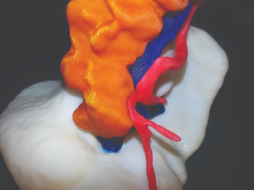
Soon after the initial trial using 3D printed medical models, the university added PolyJet™ 3D Printing technology to its prototyping laboratory, which it calls the Protolab. Now the university produces 3D printed surgical guides for every spleen and kidney operation, and about half of the pancreatic surgeries. “With its super-fine 16-micron layer printing, the Objet30 Pro provides us with highly accurate 3D printed models with the smallest of detail in VeroClear material. This is especially crucial for vascular models in order to locate the blood system,” said Stefania Marconi, researcher at the University of Pavia.
By converting CT scans into 3D printed surgical guides, the university transformed its preparation process and can now produce models for individual patient cases. These include splenectomies and donor kidney organs. The 3D printed anatomical models reduce unexpected risks and help surgeons locate access points for their instruments during laparoscopic and robotic surgeries. The university recently provided surgeons with a 3D model of a kidney to detect and procure the organ in preparation for a transplant using the robotic surgical system ‘da Vinci’.
This enabled them to navigate through the complex vascular network and recognize distances between organs, which was critical in avoiding damaging crucial vessels. As they continue to use leading technology to 3D print anatomical models for most of their operations, the University of Pavia will continue to set the standard in surgical preparation. “Thanks to the incredible accuracy, the 3D printed surgical guides have proved instantly popular amongst the surgeons,” said Auricchio. “With more of our surgeons having a physical model of the patient’s organs, it equips them with vital details before operations.”

