AI startup Medical IP has attracted a great deal of attention as a leader in the digital transformation of the Korean medical market. Since the company’s founding in 2015, fast-growing Medical IP has become a symbol of innovation in the medical market, recently receiving the first venture company recognition by Seoul National University Hospital.
Medical IP’s AI-based image processing and 3D printing technology allows hospitals, academic centers, and medical device companies to create ultra-realistic human organ models by segmenting medical images such as X-rays, CT scans, and MRIs using AI technology. Medical IP’s technology brings 2D black-and-white image data from CT and MRI scans into three-dimensional life so it can be used to create anatomy models that allow unprecedented patient treatment, prognosis, observation, training, and surgery simulation.
In the past, hospitals and academic centers have relied on animal experiments, cadavers, and legacy 3D printing technology for training. Animal and cadaver models not only raise ethical issues, but also lack the anatomical realism and repeatability that can be achieved with highlyaccurate, patient-specific 3D printed models. Similarly, legacy 3D printing technology requires multiple printed parts for each color and texture, which slows throughput, limits color and texture options, and creates a significant amount of postprocessing work.
To create ultra-realistic anatomical models in less time, Medical IP partnered with Stratasys, leader in 3D printing technology. Organ models created using Medical IP’s AI-powered segmentation technology and the Stratasys J750 are printed in full color with various textures mimic the natural shape, structure, feel, and haptic feedback as a real organ.
Medical IP's software solution maximizes the value of medical image data that has been lying dormant on hospital computers. Instead of having to visually review 2D black and white images from a monitor, Medical IP transforms the data into 3D models that practitioners can actually see and touch.
The impact of this technology on the medical field is immense.
How it works: Medical IP’s workflow
Medical IP’s segmentation technology received FDA approval and CE Mark clearance in 2019. Along with the Stratasys J750, Medical IP technology has made the 2D-to-3D printing workflow seamless.
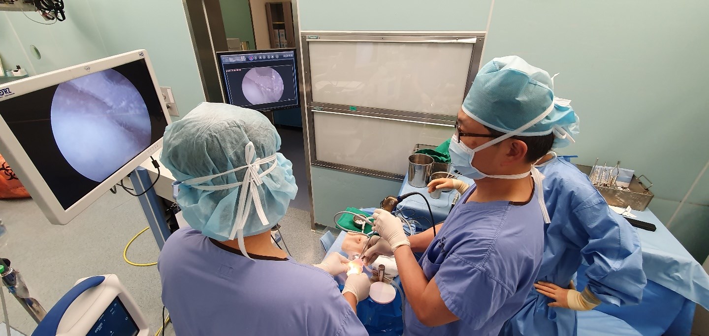
Simulated pituitary tumor removal.
Medical IP partnered with a team of clinicians to test and validate a pituitary tumor removal surgery simulator that was developed by Medical IP and printed using the Stratasys J750.
Each surgeon reported that the drilling experience was realistic as drilling into a real bone. “Blood vessels, nerves, dura, brains, etc. were very realistic and training could be carried out in an environment similar to actual surgery,” commented a surgeon.
After the testing, one participating hospital requested a long-term production plan for patientspecific, 3D printed models to be used by its pediatric surgery team for surgical planning. Unlike adults, children do not have fully grown organs, so organ shape is not consistent and it can be difficult to identify lesions. 3D printed organ models enable unprecedented case planning for improved patient outcomes.
Simulation-based training for kidney stone removal.
A medical institution sought to overcome challenges created by its traditional training methods that did not allow simultaneous urologic navigation and kidney stone grinding. To compare its existing skills training to Medical IP’s 3D-printed simulation, the institution worked with Medical IP to develop a kidney stone simulator using that could be tested with our without an endoscope for mobile training.
The medical institution immediately saw the educational value of the 3D-printed model. 3D printed kidneys allow stones to be positioned in any location, and semi-transparent material allows surgical practice to be done without an endoscope. Additionally, water can be circulated throughout the model to simulate stone grinding using both a laser and an ultrasonic to make haptic feedback ultra-realistic.
Medical IP is dedicated to find new ways to use medical AI technology to predict, treat, and manage disease. By partnering with major global medical leaders such as Stratasys, Medtronic, Johnson & Johnson, Olympus, Bard and Intuitive, Medical IP seeks to enhance AI-based medical image analysis technology and expand its core technology to new applications like 3D printing.
“AI and 3D printing can be viewed separately. But our approach is different; we are constantly looking for things that will be beneficial to patients and helpful to medical professionals. We combine AI and 3D printing technologies to create synergy, and we are planning to deliver digitalbased healthcare innovation to more medical professionals both domestically and overseas.” said Sang Joon Park, CEO of Medical IP.


