A new and vital tool is 3D printing. The hospital recently entered into a partnership with Tknika, a Research and Applied Innovation Center for Vocation Education and Training in the Basque region, and Tecnun, a specialist division of Universidad de Navarra. This partnership gives the surgical team access to more advanced 3D printing technology. “3D printing is an essential surgical tool for us,” explains Dr. Jon Zabaleta, Thoracic Surgeon at Biodonostia. “Previously, no 3D printed model we created in-house could meet the level of detail and accuracy we needed. However, thanks to our partnership with these local institutions, we now have access to advanced 3D printing technology from Stratasys that enables us to meet the demands required to create highly-accurate, patient-specific 3D models.”
Stratasys FDM 3D printing has proved particularly important when treating complex, and often life-threatening thoracic wall tumors. Located on the chest wall, thoracic tumors can cause excessive and painful swelling, or lead to trouble breathing for the patient.In a recent case, a 64-year-old man came to Dr. Zabaleta with an extremely complicated tumor on his thoracic wall. Over the course of two years, the tumor had slowly spread across multiple ribs. The man was in intense pain, concerning surgeons about his respiratory function. “Ordinarily, in a case like this, we would remove the affected ribs and correct the defect by covering the area with a titanium plate,” said Dr. Zabaleta. “These plates are a standard size, designed for men of 100 kg or women of 50 kg, and need to be altered and rotated during surgery to suit each patient’s specification. In a complicated surgery, this can add hours to the operating time.”
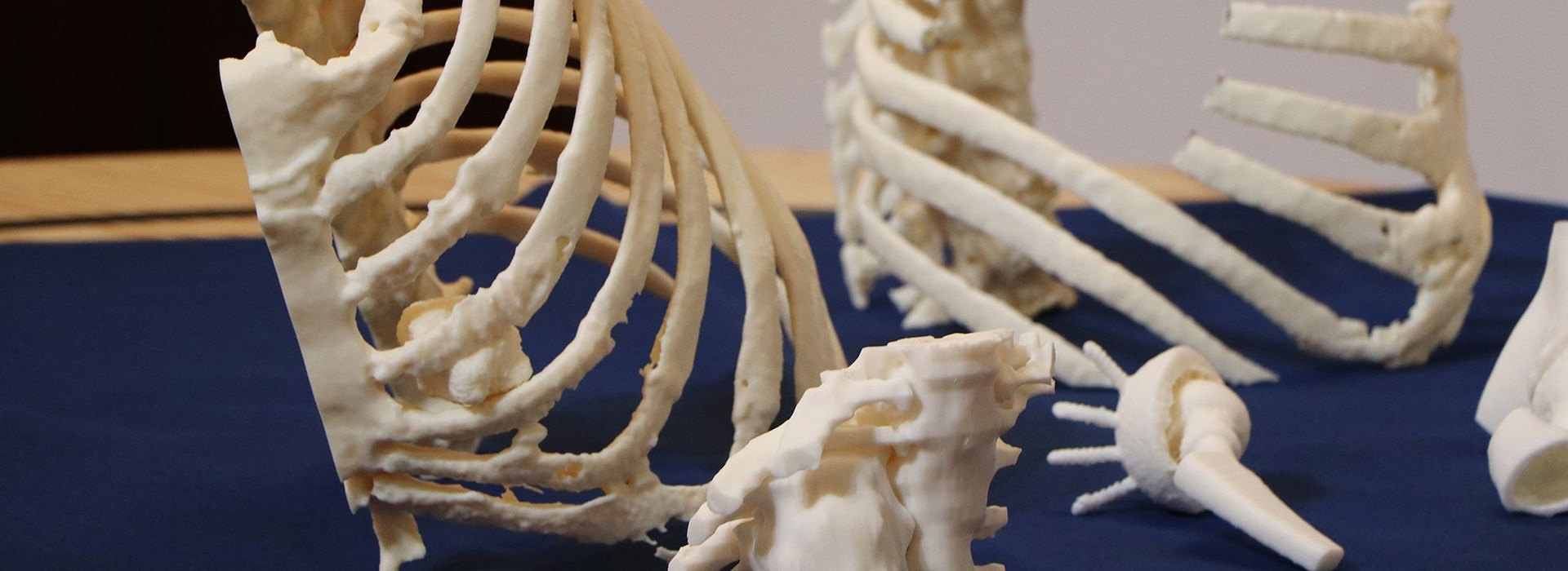
As Dr. Zabaleta explained, this case presented a complex challenge for the surgical team, since removing the tumor would require removal of more than one rib, an unusual method of treatment that increased the surgical risks. As a result, the surgeons needed to find the best way to correct the defect with the strength to protect the lungs, while maintaining flexibility and movement inthe chest. In order to explore and plan the surgery, the surgeons turned to their partnership with Tknika and Tecnun to produce an advanced, patientspecific 3D model of the patient’s thoracic wall. Together, the hospital’s partners converted a conventional CT scan of the patient into a 3D printed model using the Stratasys Fortus 450mc 3D printer and returned it to the surgical team within 24 hours.
“By creating a precise, anatomically-accurate 3D model of the thoracic wall, we were able to plan and perform the resection on the 3D model ahead of the surgery,” explains Dr. Zabaleta. “This allowed us to measure the screws and pre-bend the titanium plates in advance and helped reduce the overall operating time by 2 hours. For the patient, this meant a significant reduction in time under anesthesia, and for our hospital, freeing up time in operating rooms saves costs.”
For this condition, the surgeons required a model strong enough to replicate human bone, so the teams at Tknika and Tecnun selected FDM technology for its ability to print with engineering-grade thermoplastics. “Our partnership afforded us access to the necessary technology to produce a large and complex model that was incredibly strong, close to the real bones we would face during surgery. Without the strength of this model, we could not have prepared for the surgery in the same way,” Dr. Zabaleta explained. In addition, Dr. Zabaleta credits the 3D models with an improvement in patient-doctor communication. He used the models to explain how they would protect the lungs, which helped alleviate the patient’s anxiety ahead of a complicated operation and achieved informed consent more quickly and easily.
Additionally, this process makes the surgical consult faster and more efficient, offering the surgeon time to see more patients. Dr. Zabaleta believes that the next natural step will be for all surgical disciplines at Biodonostia to use 3D printing to prepare and plan for surgeries, as it offers the hospital the opportunity to innovate their treatment procedures and improve patient care. “The use of the 3D printed model was so essential to this case, and we are working to apply this to many other surgical disciplines across the hospital, from pancreatic tumors to airway stenosis, and these 3D printed models are already being used to help train our future surgeons,” said Dr. Zabaleta.

