Italiano
Italiano
BIOMODEX neurovascular twins.
Case Study

3D ‘twins’ give neurovascular intervention a confidence boost.

June 01, 2020

Surgeons turn to 3D printing for training.

The human brain is an incredibly complex organ supported by an equally intricate (and delicate) network of blood vessels. So for neurosurgeons, precision is critical when it comes to treatment and surgery. Every micrometer counts when faced with problems such as aneurysms and blocked arteries.

That’s why surgeons are turning to training on 3D-printed twins.

In early November 2019, a surgical team at Toronto Western Hospital, led by Dr. Vitor Mendes Pereira, a neurosurgeon and neuroradiologist at the hospital’s Krembil Brain Institute, successfully performed the first-ever robot-assisted stent placement and aneurysm coiling procedure. The robotic CorPath GRX system gave the team extra sets of arms and eyes, and unmeasured control to successfully complete the procedure and achieve a positive patient outcome.

It wasn’t as simple as dropping a robot into a surgical suite and exercising a few keystrokes. This pioneering step required extensive testing and refinement. And practice. Fortunately, Dr. Pereira and team had a readily available solution based on their use of “anatomical twins” to rehearse for other novel and complex cases.

Advances in biomechanically realistic 3D printing make practice easy and cost-effective. These anatomical twins are 3D models that mimic the structure and texture of the individual patient’s arteries and surrounding tissue. Created by medtech innovator BIOMODEX, the replicas were built using medical scans brought to life by the company’s proprietary modeling software, and Stratasys 3D printers and advanced materials.

“The realism is remarkable,” said Dr. Pereira. “We were essentially able to perform the procedure before performing the procedure and sharpen our approach. This allowed the team to enter surgery with confidence, and decrease the associated time and risk.”

The material mix has to be accurate. Precision is critical so the training or rehearsal is as close to real as possible.

BIOMODEX neurovascular twins.

Designing thousands of 3D-printed twins.

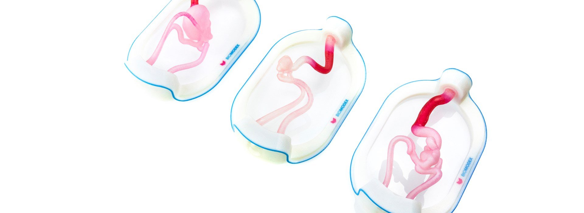
BIOMODEX develops anatomical twins to help scientists and physicians such as Dr. Pereira with patient-specific training and rehearsal. The goal is to produce models with a look, feel and experience that is true to what a doctor will encounter during a real procedure on a real patient.

The company uses multiple Stratasys J750™ printers and PolyJet™ materials to ensure the dimensional accuracy and mechanical behavior of the original anatomy. Exact down to the microscopic level, the models are paired with a portable simulation station that injects liquids to recreate blood flow.

“The material mix has to be accurate,” said Stéphane Caporusso, global VP of Operations for BIOMODEX. “Precision is critical so the training or rehearsal is as close to real as possible.”

While the use of models for medical training isn’t necessarily new, time has been a limiting factor. Historically, they’ve taken months to create — not a practical option when patients require rapid intervention. The BIOMODEX difference is using 3D printers for higher-volume, but extremely detailed production. The Stratasys printers run for 15 hours a day, every day, producing up to 3,000 models per year, per machine.

Because of the capability of the 3D printers and utility of the materials, the turnaround time from medical imaging to delivery of an anatomical twin is just a few days.

Safely introducing robots.

For Dr. Pereira, using BIOMODEX anatomical twins was critical to preparing for his first-of-its-kind, robot-assisted intervention. By using an anatomical twin, the team was able to identify, record and mitigate potential issues and ensure the stent they had selected for the patient was the correct size.

The rehearsal also provided an opportunity to integrate and coordinate the surgical assistants and CorPath GRX system. Because robotic procedures are done remotely, Dr. Pereira worked nearby while the bedside team synchronized with the robotic arm to place the stent and secure the aneurysm. The team was able to adjust their workflow for the procedure based on their learnings from the rehearsal.

“The robotic physician can’t ‘see’ what we’re doing in the same way we can,” Dr. Pereira. “So coordination and communication are key during robotic procedures.”

Once the first procedure was complete, Dr. Pereira realized the approach could have an impact far beyond the walls of this one hospital.
BIOMODEX simulation station.
BIOMODEX surgery rehearsal.

Advancing neurovascular proceduces.

Toronto Western is a teaching facility. Robotics or not, the 3D printed models from BIOMODEX and Stratasys give residents lifelike, case-specific experience as they learn and refine their skills, the type of experience they’re not able to get with cadavers.

And since the inital surgery, Dr. Pereira and his team have successfully completed several additional aneurysm interventions with the help of CorPath GRX and anatomical twins. Their success marks the first step toward his vision of remote neurovascular procedures.

“The ability to deliver rapid care through remote robotics could have a huge impact on improving patient outcomes and allow us to provide cuttingedge treatment to patients everywhere, regardless of geography,” Dr. Pereira said.

The method demonstrates the benefits and potential reach of combining robotic and 3D-printing technologies. Underserved populations could soon receive high-level care for critical procedures — with their doctor fully prepared and sitting miles away.

Download the case study

Download