While 3D printing in areas such as orthopedics, trauma and neurosurgery is widespread, Dr. Tiago Senra of the cardiovascular MRI and CT scanning department says cardiology is another story, due to complicating factors. First, blood vessels are difficult to identify in imaging tests. “To enable image diagnostics, we inject contrast in the veins or arteries,” explains Dr. Maximilliam Gospos of the cardiovascular MRI and CT scanning department. Also, because the heart is constantly beating, MRI or CT scans must be synchronized with an electrocardiogram to render static images of the veins and arteries. However, DPCI is already using 3D printing technology to help its patients. DPCI chose an Objet350 Connex because of its multi-material and highprecision capabilities. “The ability to print a prototype of an artery in rubber is very important to us,” emphasizes Senra.
“If we wish to work with a prototype that behaves similarly to an artery, it must have elastic properties. Printers that work with only rigid materials would not be functional for this use.” According to Bruno Utiyama da Silva, researcher and coordinator of the 3D printer projects at DPCI, the flexible material gives the doctors and engineers of Dante Pazzanese freedom to create new uses and solutions using the 3D printing technology. “We also use this machine in the process of developing heart pumps, which demands materials very different to those used in the blood vessel prototypes,” explains Utiyama.
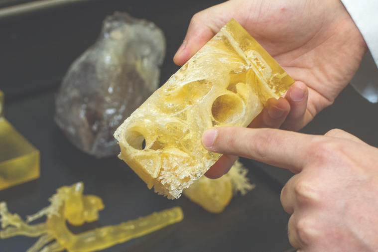
The external 3D anatomical model of the patient allows the doctors of DPCI to study the conditions of the pathology and then decide which treatment to pursue. The tests carried out on 3D printed arteries will reveal whether performing a catheterization is safe or not. With the 3D printer’s help, it is possible to study a patient’s specific blood vessel without the need for invasive procedures. To reduce the risks of patients being subjected to catheterizations, DPCI uses the 3D printer to 3D print the arteries of the patients. “The aim is to perform tests, before the catheterization, to see if it is worth performing this procedure or not,” says Senra. To perform these tests, both the patient’s artery and an “umbrella” are printed simultaneously.
The umbrella is an embolic protection device implanted in the artery before the catheter is inserted. “By adjusting the umbrella in the artery, we can be certain that the embolic protection device will, in fact, cover the area of the entire vessel,” says Gospos. “If that doesn’t happen, there is a risk the catheter will touch the walls of the vessel and dislodge embolisms. This could lead to problems for the patient.” What Gospos describes is distal embolization – when the catheter insertion dislodges small plaques from the walls of the vessels. These plaques can lodge in small vessels such as brain arteries where they could cause a stroke. A stroke can lead to serious complications, such as loss of strength in limbs and facial muscles, aphasia and sometimes death, so it’s important to correctly determine if a catheterization is right for any given patient.
The DPCI team can now prototype a patient’s vessels, study the catheter passage, analyze the risk of embolisms and determine whether to recommend catheterization. “We use 3D printed prototypes of the artery and umbrella to simulate how the catheter insertion would take place and if the vessel walls would be protected during the entire procedure,” said Senra. “The 3D printing technology helps us separate patients who are able to go through the procedure from the ones who aren’t.”

