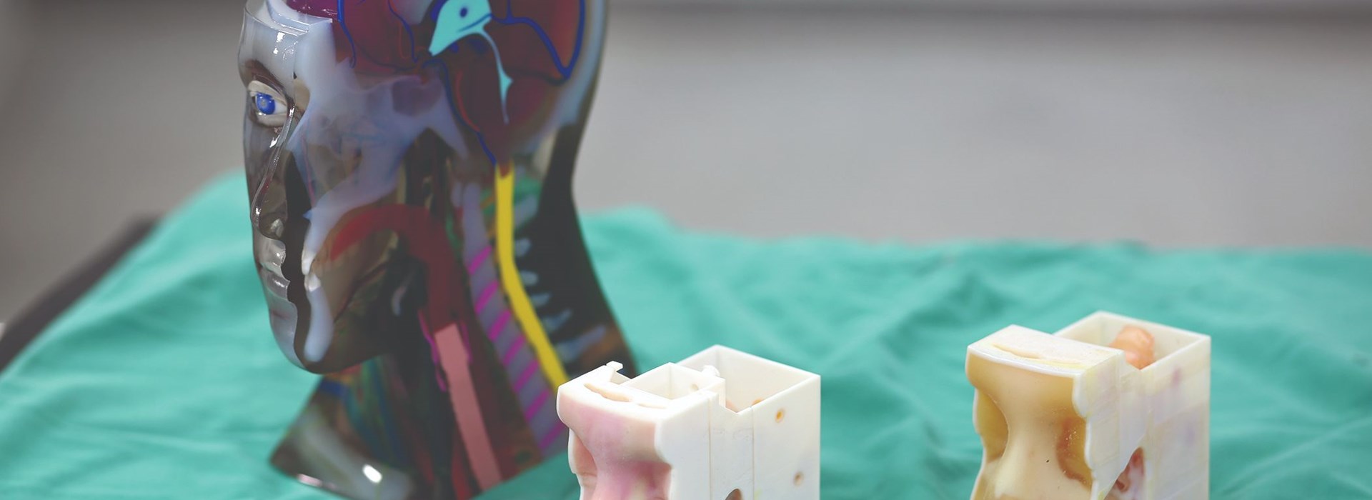
At CBMTI’s inception, the primary tools for training neurosurgeons were mentoring on live human cases, cadaver dissection and computer simulations. In a groundbreaking step to enhance training, the center adopted 3D printing. Its first 3D printer made spatially accurate models in a single material but did not mimic human pathology without a costly, time-consuming post process. This changed when they acquired PolyJet 3D printing technology.
“Once we got the Stratasys multi-material printer, we were able to print models that could, for instance, mimic the texture of the nose, the linings, and the harder tissue at the back of the nose. We found this very useful, especially in teaching trainees how to handle different materials,” said Vicknes Waran, MD, director of CBMTI.
A recent program was developed to train surgeons on ear, nose and throat surgeries in collaboration with Professor Prepageran Narayanan from theUniversity of Malaysia.“When you use a 3D model with a tumor or lesion, it is very important to have color. Only if you see the color separation do you know that you’re in the right plane,” said Narayanan. “Now you can use a model based on a patient’s pathology to simulate the entire surgery before the surgery itself.”
Interest has significantly increased since CBMTI invested in 3D printing and the company has increased production capacity by 40 percent with its 3D printers. A team of 20 medical clinicians, rapid-prototyping engineers, computer programmers, and electrical engineers work together on their main 3D printing lines of business: creating prototypes for university research, developing custom titanium implants and manufacturing custom simulators for surgical training.
“Researchers’ interest in our models has increased a hundredfold since we began using these printers,” said Balakrishnan. “Stratasys printers are the ideal platform for innovation. We have gone from being only able to mold titanium plates for cranial implants to being able to create bio-models with pathology from actual patient imaging data.
CBMTI now 3D prints detailed multi-material models that mimic real anatomy, even down to a specific patient’s tumor. With access to advanced multi-material 3D printing, CBMTI can fabricate models that feature different textures and densities over surfaces and throughout interiors, just as human body parts do.“The [Stratasys] J750 allows us to create models with both texture and color variations that mimic actual tissue handling and appearance better for these complex models,” said Dr. Waran. “With the Connex, we can simulate realistic layers of human tissue like skin, bone, dura, brain, and tumors within the printed model for surgical simulations.”
CBMTI develops its training courses in partnership with leaders in various fields. Together, they identify a patient with the anatomy and pathology they wish to train physicians to treat. CBMTI engineers then convert the patient’s CT and MRI scans into digital design files and select materials that best match the physical, tactile and color characteristics of the target anatomy. CBMTI has even found ways to use support material, typically removed from the final model, to enhance clinical realism.“We have also incorporated features such as fluid dynamics so we can simulate endoscopic neurosurgical procedures,” said Yuwaraj Kumar Balakrishnan, CBMTI chief operations officer.
“We find surgeons who train on these models are much better prepared in terms of dealing with complex surgeries, simply because they are able to train and retrain on the models until they perfect the procedure.”


