USA & Canada
USA & Canada
3D printed phantoms (hero image)
Case Study

Creating realistic imaging simulations with 3D printed phantoms.

April 01, 2020

The Children's Hospital and Medical Center in Omaha is using 3D printed phantoms to create a safer environment for patients.

3D printing phantoms at the Children's Hospital and Medical Center in Omaha.

Seeking lifelike, low-risk solutions.

With expertise in more than 50 pediatric specialties, Children’s Hospital & Medical Center in Omaha has the largest group of specialty pediatric physicians in Nebraska and serves children throughout a five-state region. Children’s Criss Heart Center was ranked among the best pediatric cardiology programs in the nation by U.S. News & World Report in 2019. The center offers comprehensive cardiac care programs for both children and adults with congenital heart disease, including pediatric heart transplantation.


Children’s state-of-the-art cardiac catheterization lab (cath lab) features a robotic 3D imaging system manufactured by Siemens Healthineers, called the ARTIS pheno — making it the first pediatric cath lab in the world to use this camera.


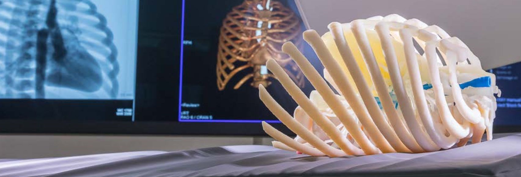
What makes this technology so revolutionary is its fusion functionality, which allows physicians to align an active fluoroscopic image to a previously obtained MRI or CT scan to develop a 3D roadmap for the cardiac catheterization procedure.


With the introduction of the ARTIS pheno, Children’s wanted to ensure that cardiologists and the cardiovascular interventional radiographers had the most realistic training possible. “To test and train, you want to make the scenario as lifelike as possible. That way your patients are not receiving additional radiation or contrast once they’re on the table because you’ve already practiced your approach,” explained Gabe Linke B.S, ARRT (R)(MR), Advanced Imaging Program Coordinator at Children’s.

 
The problem was that the most realistic training option at the time was to use live patients. However, subjecting such young and fragile patients to more radiation than necessary was out of the question. Another option for interfacing with and testing the new technology was to scan a cell phone and practice fusing the images. But cell phones are not representative of actual human anatomy, so the team sought out a better solution.

We use 3D printing to improve patient outcomes on any scale. And when there's not a solution to a patient-specific problem or a process that needs to be improved, 3D printing has been a game changer.

Creating a realistic scanning simulation.

The team at Children’s decided that creating and using a phantom — an object designed to mimic human anatomy when scanned by medical imaging equipment — would be the best solution for refining skills on the ARTIS pheno imaging system.

To develop a phantom that would provide the most realistic training simulation, cardiologists, radiologists and the 3D imaging team worked together to design the digital files. At the time they only had a single material printer on campus and they knew they needed multiple materials to achieve a realistic phantom. So they reached out to Dr. Jorge Zuniga and the University of Nebraska Omaha’s biomechanics research team. Together, using the Stratasys® Objet260 Connex3™ 3D printer, they were able to 3D print a multi-material phantom that replicated the look, feel and function of real patient anatomy. The phantom was based off a pre-existing scan of a coarctation — a common congenital heart condition — and featured a rib cage and airway as well as a hollow heart cavity and vessels that would allow contrast to be injected. This ensured that those training with the phantom and ARTIS pheno would achieve the most realistic scanning simulation.

With this new 3D printed phantom in use, Children’s Hospital & Medical Center started seeing immediate results. “We put out a survey to all the technologists that used it in training and 85 percent of them said they preferred having a contrast-filled phantom to work with,” said Blair Kauzlarich B.S, ARRT (R) CI, Advanced Imaging Program Coordinator at Children’s. This allowed cardiovascular interventional radiographers to refine their skills and processes with the imaging equipment before a patient entered the cath lab.

Children’s has also noticed that the reduction in time spent in the cath lab and the operating room has supported their position to have an in-house 3D Print Lab. “When you’re saving that time, and are better prepared for cases, it’s easy to understand the need for this technology,” explained Linke. “And the benefit of having a realistic lifelike simulation was invaluable to our team. How do you put a dollar amount on having a safer environment for your patients?” added Kauzlarich. Overall, the phantom greatly improved the ARTIS pheno system’s launch at Children’s.

...the benefit of having a realistic lifelike simulation was invaluable to our team. How do you put a dollar amount on having a safer environment for your patients?

Improving patient outcomes.

Since the creation of Children’s first 3D printed phantom, it has added the Stratasys J750™ multi-material printer to its 3D imaging arsenal. 3D printing has also become a multifaceted effort with a dynamic team led by radiology that extends to other subspecialties throughout the hospital. “We use 3D printing to improve patient outcomes on any scale,” stated Linke. “And when there’s not a solution to a patient-specific problem or a process needs to be improved, 3D printing has been a game changer.”
3D printed ribcage (side)

Download the case study

Download