As the director of the structural heart team at Kaleida Health’s Gates Vascular Institute (GVI) in Buffalo, New York, I perform minimally invasive, endovascular surgeries to repair structural defects of the heart, whether a defective valve or other holes or defects. I specialize in transcatheter aortic, mitral, and pulmonic valve replacements, transcatheter mitral clip procedures, as well as closures on patent foramen ovales (PFOs), atrial septal defects (ASDs) and ventricular septal defects (VSDs) among other structural heart treatments. Rather than cracking open the chest cavity to access the heart, I perform these as endovascular procedures, using the body’s blood vessels as the conduits through which medical devices are delivered. Although less risky than open-heart surgery, endovascular procedures are complex, so the ability to practice procedures and deploy devices in realistic models is extremely valuable.
The patient-specific vascular flow models are created by biomedical engineers. They take CT scans, or medical images, of the patient’s heart and convert them into the type of file that is uploaded into a 3D printer to create an accurate multi-dimensional anatomy. This conversion process can be performed in a matter of hours and we are looking for ways to accelerate it.
Using 3D printers is currently the best way to replicate patient-specific anatomy. We 3D print vascular flow models, sometimes called vascular phantoms, to simulate mechanical properties of blood vessels, tissue, and calcifications which allows us to “see” if navigation and vessel perforation is possible. The Jacobs Institute (JI) a non-profit medical device innovation center, in partnership with the University at Buffalo’s Toshiba Stroke and Vascular Research Center (TSVRC), the GVI, and Stratasys, uses a Stratasys Objet500 Connex3 3D Printer to produce the multi-material vascular models with rubberlike, polyurethane blend materials that have different material properties. The models simulate structural heart as well as neuro-, cardio-, and peripheral-vascular procedures. Biomedical engineers prepare the patient-specific models using CT scans or other medical images which are converted to 3D printable files.
Advantages of 3D Printed Models Over Alternative Methods
Before 3D printed vascular flow models, planning for surgery essentially entailed looking at 3D reconstructions from patients’ CT scans on the computer and trying to imagine how the devices would fit. Because it was difficult to extrapolate all of the necessary information from these 3D reconstructions, adaptations had to be made on the fly during the procedures themselves. This both lengthened the procedure time and added another element of uncertainty. While cadavers offer the opportunity to practice deploying devices, they do not mirror the anatomy of specific patients, nor do they feel like living vessels, as the vessels are formalin fixed.
Using the 3D printed heart model for surgical planning
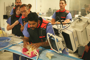
Simulated Cath Lab Setup To Pre-Plan
Now 3D printed models can be hooked up to a “pump” that simulates blood flow and allows for physicians to experiment with different routes for endovascular procedures. Using 3D printed vascular models to plan complex surgeries contributes to procedure success and better outcomes for high-risk patients, especially when I am using a new device and/or an off-label device. As each patient and situation is unique, being able to test a device on a three-dimensional model prior to a complex procedure allows physicians to understand the potential interactions of the device with the patient’s tissue, as well as how the device will sit in the space. All of this information allows me to individualize surgical strategies.
Click here to stay on top of 3D printing innovation in healthcare
What I Learned From a Pulmonic Valve-in-valve Replacement
In a recent pulmonic valve-in-valve replacement, based on computer based imaging, I was planning to proceed from the right femoral vein through the right atrium, right ventricle and then through the RV outflow tract atrium into the pulmonic valve. However, during the dry-run on the 3D printed model of the patient’s heart, the device buckled in the right atrium when we tried this course. Based on this feedback, we adapted our plan and used a stiffer wire so it provided a tighter rail.
In terms of future improvements, the models need to be more predictable with better tissue characteristics so they respond to devices as real tissue would. It is probable that better imaging will lead to better models and that we will be able to make more accurate models using multiple modalities (CT, Echo, MRI) of the structures.
While the models are extremely helpful planning and practicing complex procedures, they do not need to be used for routine day-to-day cases as they are not without cost both in terms of materials and time. The collaboration between JI and Stratasys, allowing our physicians access to advanced multi-material 3D printing technology, is invaluable for complex cases and offers exciting opportunities for learning as medical devices and endovascular procedures become more intricate.
Learn how to plan and implement your own 3D printed surgical modelling program with this complimentary white paper.


