3D printing is transforming surgical planning and enabling the delivery of personalized medicine to infants with complex congenital heart disease (CHD). Five years ago, the cardiovascular team at SSM Health Cardinal Glennon Children’s Hospital, a pediatric medical center in St. Louis, Missouri, embraced 3D printing as a solution for better surgical planning for these patients. While congenital heart defects are the most common birth defects worldwide according to the American Heart Association, affecting eight out of every 1,000 babies born in the United States, fortunately only one in four requires surgery during the first year of life. As a pediatric referral center, SSM Health Cardinal Glennon Children’s Hospital sees some of the most complex patients, requiring repair within days or weeks of birth on hearts the size of a walnut.
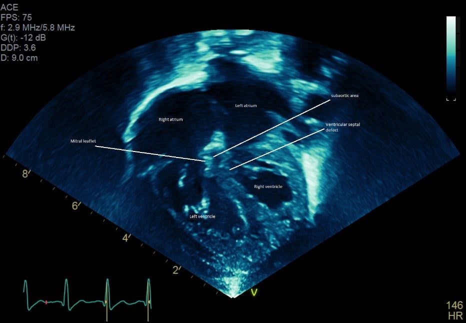
One such patient was an infant diagnosed in utero via fetal ultrasound with an unusual form of transposition of the great arteries. Deoxygenated blue blood entered the right atrium which connected to the left ventricle and then the aorta, and oxygenated red blood entered the left atrium which connected to the right ventricle and then the pulmonary artery. The patient also had a very large ventricular septal defect (VSD) connecting both ventricles and severe narrowing between the left ventricle and the aorta. It was apparent early on that the patient was becoming fairly blue as deoxygenated blood was being directed towards the aorta. A balloon atrial septostomy performed in the first few days of life resulted in some improvement. However, significant tachycardia persisted. It became clear that the degree of subaortic narrowing was more severe than originally anticipated, and this resulted in significantly depressed left ventricular function. Something needed to be done soon.
The cardiothoracic surgical team led by Charles Huddleston, M.D., and members of the cardiac medical team including Wilson King, M.D., agreed that surgery was required. Due to the rarity of this condition, there was controversy over how to repair the infant’s heart. A neonatal atrial switch seemed to be the best strategy. This is a challenging operation that redirects the oxygenated blood to the aorta and deoxygenated blood to the pulmonary arteries by creating intraatrial baffles. Addressing the subaortic stenosis was extremely problematic, as it appeared that much of the obstruction was caused by tissue that was extremely close to the mitral valve. Damaging the mitral valve would result in significant problems for the patient. “After reviewing the echocardiogram, many of our cardiologists as well as the referring cardiologists were convinced it was part of the structural integrity of the mitral valve and resecting it would mean replacement of that valve and subjecting the child to lifelong disease. Others thought it was accessory tissue not critical to mitral valve function. From the imaging studies alone, it was impossible to tell, limiting our ability to confidently develop a surgical plan,” said Dr. Huddleston (figures 1,2,3,4). For complex cases like this, the Cardinal Glennon surgical team relies on 3D printing.
Using an MRI dataset, the team 3D printed a model of the infant’s heart using a Stratasys J750™ 3D printer. “It’s hard to judge what to do looking at 2D images on the screen. It is difficult to capture all the information in one plane even with a 3D reconstruction on a 2D screen. Spatial relationships are a lot easier to conceptualize with a 3D model. With the 3D model it became evident how to redirect the blood flow to the right places to provide oxygen to the body,” said Dr. King. Dr. Huddleston added, “The 3D model helped us identify the anatomy clearly and provided us with the confidence that the accessory tissue causing the subaortic stenosis was not an integral part of the mitral valve. This allowed us to go into the procedure assured that the tissue could be removed safely without damaging the mitral valve located near it.” Dr. Huddleston summed up his experience, “The 3D model was instrumental in determining the optimal surgical approach. It allowed us to go into the case with a solid plan based on the best information we could obtain.” With the help of the 3D printed heart model, the neonatal atrial switch, VSD closure, and subaortic stenosis resection was successfully performed in a 20-day-old infant, and the mitral valve function remained intact. The cardiac function gradually improved, and the patient’s heart rate normalized. He is expected to have an excellent long-term prognosis.
")