Traditionally, surgeons plan operations based on the CT and MRI images of patients’ conditions. While these images can illustrate a patient’s organ from different angles, they might not show all injuries that could cause possible complications. The Objet500 Connex3 solves this problem: Sugimoto 3D prints full-sized models of patients’ internal organs. “The multi-color and multi-material bio-models help surgeons uncover hidden tissues and blood vessels that may be blocked by larger organs in the 2D scans,” says Sugimoto. “Clearer perspectives can now be visualized prior to operation and more accurate treatments can be planned as a result.”
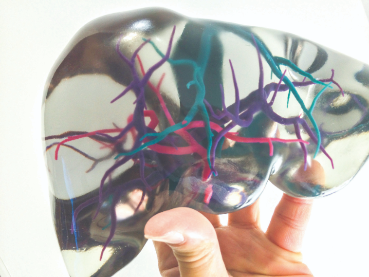
Surgeons examine the models’ different angles – and even mark up the models – to plan surgical procedures, drastically reducing the operation time. “Customized bio-models are created with the most accurate information available, including cancer cells’ exact location, and how each vein and artery is attached to the organ or tumor,” explains Sugimoto. “These models are used to identify the appropriate treatments and simulate surgical procedures. In addition, these 3D printed models are used as tools when communicating the operation process to patients, thus providing a better understanding of the procedure.” To replicate a patient’s liver for pre-surgical examination and preparation, Sugimoto and his team converted the patient’s CT and MRI data into compatible digital files, then 3D printed the liver using different materials: transparent resin for the organ and different colored materials for the blood vessels, portal vein and hepatic artery. With the model, surgeons could easily distinguish one from the other, better understand the patient’s condition and plan appropriate treatment such as possible medication or necessary segmental resection.
Sugimoto and his team further expanded bio-texture modeling for education and training purposes. For instance, groups of closely related organs are 3D printed for medical students to learn their structures and relations through veins and tissues. “Internal organs do not exist as independent parts in the human body. They are interrelated and surrounded by tissues, fat and body fluids, so it is essential for students to understand how organs are linked with one another, what and how they need to be aware of before performing operations,” Sugimoto says. Medical trainees can even practice resecting and stitching organs on surgical navigation systems using the 3D printed models. For example, Sugimoto designed a full-sized kidney model that was buried among adipose tissue, or fat, which was reproduced by 3D printing soft material to give similar texture. Students need to feel their way through and sort out the tissue before getting to the kidney and operating on it.
In other training sessions, students must bind a blood vessel and operate on the organ within a time limit to avoid endangering patient’s health. 3D models can help students understand which blood vessel they need to bind or where to insert a scalpel. “Bio-texture modeling by 3D printer is supporting doctors not just in surgical rehearsals, but also for clinical diagnosis. It has pushed the techno-medical boundary further and we are encouraged to keep experimenting with other possible applications of multi-material 3D printing in the medical field,” says Sugimoto.

